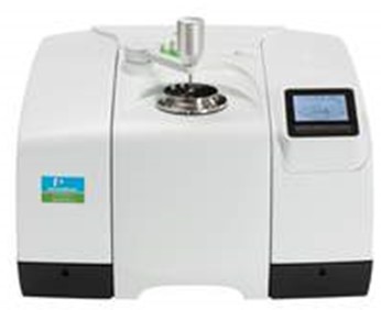
Optical Microscopy
Phi Nanoscience Center (PNSC)
1. What is Optical Microscopy?
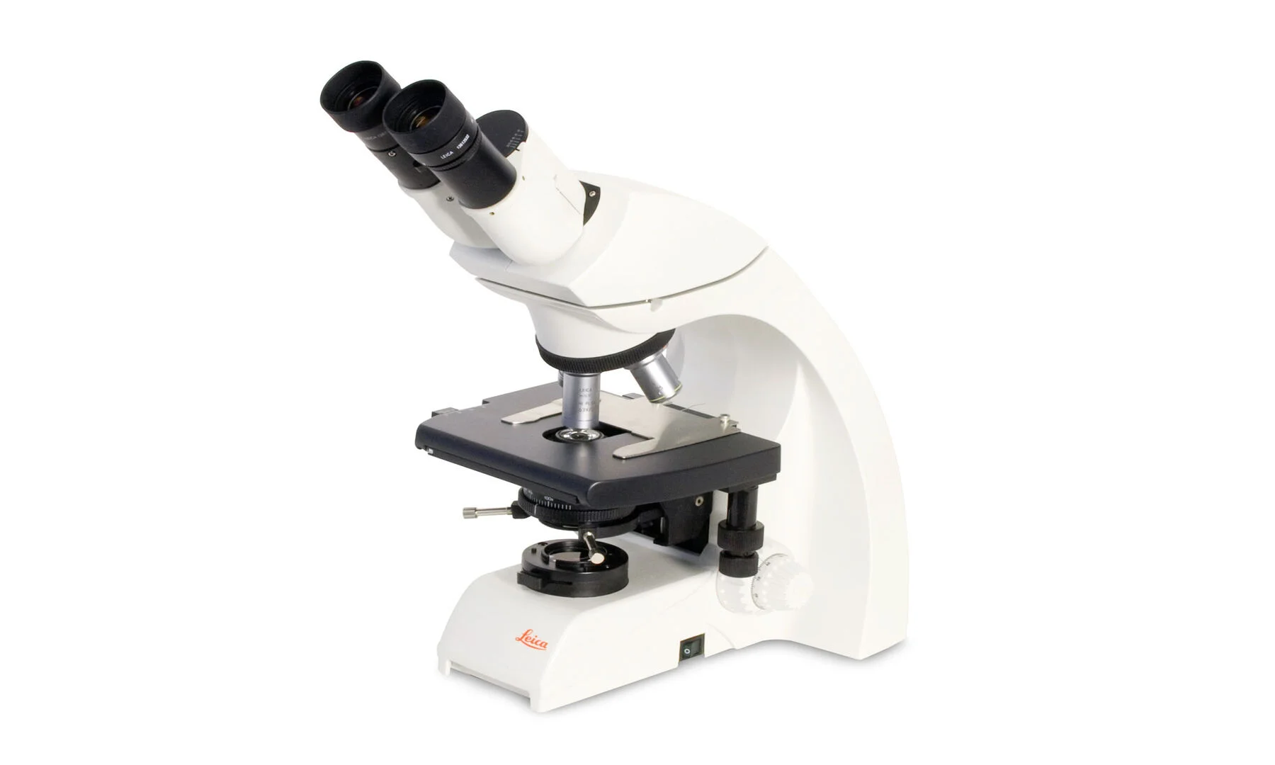
Optical microscopy (also called light microscopy) is a fundamental imaging technique that uses visible light and lenses to magnify images of small samples. It is one of the simplest and most widely used characterization tools [1].
Key Applications in Nanomaterials Research:
- Quick, initial visualization of the overall sample (e.g., are the particles dispersed or agglomerated?).
- Examining the surface of large samples, coatings, and films.
- Optical quality control for transparent or semi-transparent materials.
- Identifying large features (e.g., > 1 µm) before higher-resolution SEM imaging.
2. Principle of Operation (Simplified)
- Step 1: Light from a source (e.g., halogen lamp or LED) is focused onto the sample through an objective lens.
- Step 2: The light interacts with the sample (reflected, transmitted, or absorbed).
- Step 3: The light from the sample is collected by the same or a different objective lens.
- Step 4: The image is magnified through an eyepiece and/or captured by a digital camera.
- Step 5: The final image can be viewed in real-time or saved as a digital file [2].
Modes of Operation:
- Brightfield: Standard mode; sample is illuminated from below (for transparent samples) or above (for opaque samples).
- Darkfield: Only light scattered by the sample reaches the objective; edges and defects appear bright on a dark background.
- Polarized Light: Uses polarized light to study birefringent materials (e.g., crystals, some polymers).
3. Information You Will Receive in Your Report
- Digital Images: High-resolution images at various magnifications (e.g., 50x, 100x, 200x, 500x, 1000x).
- Scale Bar: A calibrated scale bar on each image.
- Image Details: Magnification, mode (brightfield, darkfield, etc.), and any image processing applied.
- Observation Report: A brief description of what is observed (e.g., "Spherical particles, some agglomeration, size range 1-5 µm").
4. Sample Preparation Guide
Sample Type
Preparation Method
Powder
Disperse a small amount of dry powder on a glass slide, cover with a coverslip.
Solid / Pellet / Thin Film
Place the sample directly on the stage. For thin films, no preparation needed.
Liquid Suspension
Place a drop of the suspension on a glass slide and cover with a coverslip.
Important Notes:
- The sample must be opaque or suitably prepared for light transmission/reflection.
- For transparent samples (e.g., polymers, thin films), no special preparation is needed.
5. Understanding Your Results (Guide to Interpretation)
- Spatial Resolution: The minimum resolvable distance (limited by the wavelength of light). Practical resolution for most optical microscopes is ~0.2-0.5 µm (200-500 nm).
- Magnification: The ratio of the image size to the actual sample size (e.g., 1000x means the image appears 1000 times larger than the actual object).
- Color: Provides information about the sample's interaction with light (e.g., white powder, yellow solution, colored thin film interference patterns).
Limitations: Optical microscopy cannot resolve individual nanoparticles (< 100 nm). For that, you need electron microscopy (SEM or TEM).
6. Frequently Asked Questions (FAQ)
- What is the resolution limit? Approximately 0.2 µm (200 nm) for the best visible-light microscopes. You cannot see individual nanoparticles (which are < 100 nm) with optical microscopy.
- What is the maximum magnification? Typically 1000-1500x with oil immersion lenses; beyond that, the image is empty magnification (no additional detail).
- Can you measure particle size? Yes, using image analysis software (e.g., ImageJ). The measurable size range is > 0.5 µm.
- How much sample do you need? A very small amount (milligrams of powder or a few µL of liquid).
- How long will the analysis take? 1-2 hours from sample receipt.
7. References
- [1] Murphy, D. B., & Davidson, M. W. (2012). Fundamentals of Light Microscopy and Electronic Imaging (2nd ed.). Wiley-Blackwell.
- [2] Abramowitz, M., & Davidson, M. W. (2003). Introduction to Microscopy. Olympus America Inc.
- Internal Source: Phi Nanoscience Center (PNSC) offers optical microscopy for initial sample screening and quality control.
8. Request This Test
To request Optical Microscopy analysis or any of our other services, please complete the Sample Testing Request Form.
No reviews yet. Be the first to share your experience.