X-ray Photoelectron Spectroscopy (XPS)
Phi Nanoscience Center (PNSC)
1. What is XPS?
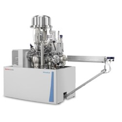
X-ray Photoelectron Spectroscopy (XPS), also known as Electron Spectroscopy for Chemical Analysis (ESCA), is a powerful, non-destructive surface analysis technique used to determine the elemental composition, chemical state, and electronic structure of materials .
XPS is uniquely sensitive to the top 1-12 nm of a sample surface, making it indispensable for studying surface modifications, thin films, coatings, and nanomaterials where surface chemistry governs performance .
Key Applications in Nanomaterials Research:
- Elemental Identification (Qualitative & Quantitative): Identifying all elements present (except H, He) and their relative concentrations.
- Chemical State Analysis (Oxidation States): Distinguishing between different chemical states of the same element (e.g., Fe²⁺ vs. Fe³⁺, metallic Ag vs. Ag⁺ in Ag₂O, Ce³⁺ vs. Ce⁴⁺ in ceria nanoparticles) .
- Surface Composition Verification: Confirming surface functionalization (e.g., PEGylation, amination) and detecting surface contaminants.
- Depth Profiling (Ion Etching): Determining how composition changes with depth (e.g., coating thickness, oxide layer composition).
- Failure Analysis: Identifying the cause of poor adhesion, corrosion, or degradation by analyzing the outermost surface layers .
2. Principle of Operation (Photoelectric Effect)
- Step 1 (X-ray Irradiation): The sample is placed in an ultra-high vacuum (UHV) chamber and irradiated with a monochromatic beam of soft X-rays (typically Al Kα at 1486.7 eV) .
- Step 2 (Photoelectron Ejection): When an X-ray photon strikes an atom near the surface, it transfers its energy to a core-level electron. If the photon energy (hv) is greater than the electron's binding energy (BE), the electron is ejected as a photoelectron.
- Step 3 (Kinetic Energy Measurement): The spectrometer measures the kinetic energy (KE) of the ejected photoelectron.
- Step 4 (Binding Energy Calculation): The instrument calculates the electron's Binding Energy (BE) using the equation: BE = hv - KE - φ, where φ is the work function of the spectrometer .
- Step 5 (Spectrum Generation): A spectrum is produced (Intensity vs. Binding Energy). Each element has a unique set of characteristic peaks at specific BE values, serving as an elemental "fingerprint." The exact BE of a peak is sensitive to the atom's chemical environment (oxidation state, bonding partners), resulting in chemical shifts .
3. Information You Will Receive in Your Report
Information
How It Benefits Your Research
Survey Spectrum (0-1200 eV): A wide scan identifying all elements present on the surface.
Provides a quick overview of surface composition and detects contaminants.
High-Resolution (HR) Spectra: Detailed scans of specific element peaks (e.g., C 1s, O 1s, Ag 3d, Ce 3d).
Used for chemical state analysis and peak fitting.
Quantitative Atomic Composition (at%): Relative concentration of each element detected (excluding H, He).
Tells you the exact surface stoichiometry.
Chemical State Assignment: Identification of specific chemical states (e.g., 15% metallic Ag⁰, 85% Ag⁺ in Ag₂O).
Confirms synthesis success (e.g., Ce³⁺/Ce⁴⁺ ratio in ceria) and detects surface oxidation.
Peak-Fitted Spectra: High-resolution spectra with individual component peaks fitted to the raw data.
Provides quantitative data for each chemical state.
Depth Profile (Optional): A plot of composition (at%) vs. sputter time (depth).
Reveals how surface chemistry changes with depth (e.g., coating thickness, oxide layer structure).
4. Sample Preparation Guide
Proper sample preparation is critical for obtaining high-quality, reliable XPS data. XPS requires an ultra-high vacuum (UHV), so samples must be dry, solid, and vacuum-compatible.
General Requirements:
Requirement
Detail
Maximum Sample Size
Typically < 15 mm in any lateral direction. Maximum height is 3-4 mm .
Sample Type
Powders, thin sheets, wafers, films, fibers, and some viscous oils (on a substrate) .
Vacuum Compatibility
Must be dry, non-volatile, and stable under UHV. No water, residual solvents, or oils .
Outgassing
Powders and polymers may require longer pumping or gentle heating (outside the chamber) to remove adsorbed gases.
Sample-Specific Preparation:
Sample Type
Preparation Method
Important Notes
Powder
Press the powder into a smooth pellet (5-10 mg) using a clean pellet press. Mount the pellet on the sample holder using double-sided tape or conductive silver paint .
For very fine powders, pressing onto indium foil can improve conductivity.
Thin Film / Coated Wafer
Mount the wafer directly on the sample holder using clips or conductive tape. For non-conductive films, ensure good contact to minimize charging.
Clean the sample surface with a gentle gas jet (N₂ or Ar) to remove loose dust .
Solid / Metal / Ceramic
Clean the surface with a suitable solvent (e.g., IPA, acetone) to remove surface grease. Mount using clips. For bulk analysis, expose a fresh surface by scraping with a clean razor blade or fracturing .
Analyze the "as-received" surface first to identify contaminants, then analyze a fresh surface for bulk chemistry.
Fibers / Powders / Fragile Samples
Wrap in clean aluminum foil or mount on double-sided carbon tape.
Ensure sample is secure and will not fall off in UHV.
Magnetic Samples
Can be analyzed but may require demagnetization. Must be very small (a few mm) to minimize interference with the electron lenses. Notify us in advance.
Degaussing may alter the surface chemistry of some materials.
Critical Note: Do NOT analyze the exact area of interest by SEM-EDX prior to XPS. The electron beam from SEM can cause carbon contamination and degrade the true surface chemistry, leading to inaccurate XPS results .
5. Understanding Your Results (Guide to Interpretation)
XPS data interpretation is a multi-step process requiring expertise and reference data.
Survey Spectrum:
- Peak Identification: Each element produces a series of peaks (e.g., Ag 3d, O 1s, C 1s). Identify peaks using a reference table or database. All peaks except H and He are detectable.
- Contaminants: Carbon (C 1s) and oxygen (O 1s) are common surface contaminants (adventitious carbon). The C 1s peak at 284.8 eV is often used as a binding energy reference for charge correction .
High-Resolution (HR) Spectra & Chemical Shifts:
- Chemical Shift: Changes in oxidation state, bonding, or coordination alter the binding energy of core-level electrons, shifting the peak position.
- Metallic State (e.g., Ag⁰): Lower binding energy.
- Oxidized State (e.g., Ag⁺ in Ag₂O): Higher binding energy (positive chemical shift) .
- Peak Fitting (Deconvolution): Overlapping peaks from different chemical states are resolved by computer fitting using mixed Gaussian-Lorentzian functions . This requires careful model development based on chemical constraints (e.g., spin-orbit splitting, expected oxidation states) .
Example Fitting Parameters (C 1s for Carbon-Containing Materials):
Binding Energy (eV)
Chemical State Assignment
FWHM (eV)
284.8
C-C / C=C (Adventitious Carbon – Reference)
1.0-1.2
286.0 – 286.5
C-O (Hydroxyl, Ether)
1.2-1.5
287.0 – 288.0
C=O (Carbonyl)
1.2-1.5
288.5 – 289.5
O=C-O (Carboxyl, Ester)
1.2-1.5
290.0 – 291.0
Carbonate (CO₃²⁻) or π-π* shake-up satellite
1.2-1.8
Practical Interpretation for Nanomaterials Research:
- Successful Nanoparticle Synthesis: For silver nanoparticles (AgNPs), a dominant Ag 3d5/2 peak at ~368.2 eV indicates metallic Ag⁰. A shift to higher BE (> 368.5 eV) and a broad, poorly defined peak indicates surface oxidation (Ag⁺) .
- Oxide Stoichiometry: For ceria (CeO₂) nanoparticles, the Ce 3d spectrum has a complex, multi-peak structure. The ratio of Ce³⁺ to Ce⁴⁺ peaks indicates the concentration of oxygen vacancies.
- Surface Functionalization: Successful attachment of a thiol (R-SH) to a gold nanoparticle surface would be confirmed by the appearance of an S 2p peak.
- Contamination: Unexpected elements (e.g., Si, Na, Cl, Ca) may indicate contamination from synthesis (unreacted precursors) or handling.
6. Frequently Asked Questions (FAQ)
Question
Answer
What is the sampling depth of XPS?
Typically 1-12 nm (approximately 3-10 atomic layers). It depends on the kinetic energy of the photoelectron and the material.
What elements can XPS detect?
All elements except Hydrogen (H) and Helium (He) .
What are typical detection limits?
0.1-1.0 atomic% (1000-10,000 ppm) for most elements .
Is XPS quantitative?
Yes, XPS is inherently quantitative (not just semi-quantitative) when using correctly determined relative sensitivity factors (RSFs). Absolute accuracy is typically ±10-20% for major components.
What is the lateral resolution?
Standard XPS: 10-200 µm. Small-spot XPS: < 10 µm. Imaging XPS (XPSI) can achieve sub-5 µm resolution.
What is the binding energy (BE) scale calibration?
The BE scale is calibrated using pure metal standards: Au 4f7/2 (84.0 eV), Ag 3d5/2 (368.3 eV), Cu 2p3/2 (932.7 eV) .
Why is charge correction necessary?
Insulating samples can develop a positive surface charge as photoelectrons are emitted, shifting all peaks to higher BE. The C 1s peak at 284.8 eV (adventitious carbon) is the most common internal reference .
What is the difference between XPS and EDX?
XPS is a surface technique (1-12 nm depth) that provides chemical state information (e.g., oxidation state). EDX is a bulk technique (0.5-2 µm depth) that provides only elemental identification .
Can XPS be used for depth profiling?
Yes, by alternating ion sputtering (e.g., with Ar⁺ ions) with XPS analysis. This is destructive to the sample .
7. References
- [1] MtoZ Biolabs. (2025). X-ray Photoelectron Spectroscopy (XPS) Testing Service .
- [2] The XPS Research Institute (XRI). (2025). Sample Preparation .
- [3] Applied Surface Science. (2024). Correlation analysis in X-ray photoemission spectroscopy .
- [4] University of Warwick. (2024). X-Ray Photoelectron Spectroscopy (XPS) .
- [5] NEWARE. (2024). XPS High-Resolution Spectra: Tool for Chemical State Analysis .
- [6] Journal of Applied Surface Science. (2022). Review on surface-characterization applications of X-ray photoelectron spectroscopy (XPS) .
- [7] The XPS Library. (2026). Sample Preparation .
8. Request This Test
To request XPS analysis or any of our other services, please complete the Sample Testing Request Form using the link below. We will contact you within 24 hours to discuss your sample, specific elements of interest, required chemical states, and any special handling requirements.